Successfully identified several factors as a result of continuing studies
to understand the mechanism of polarized transport in cells
Professor Satoh has been studying the molecular mechanism of polarized transport using Drosophila photoreceptor cells of as a model system. The molecular mechanism of polarized transport still has many uncertainties, which have been gradually clarified through recent research.
“Polarized transport” refers to vesicular transport in polarized cells, such as neurons, epithelial cells, and photoreceptor cells. (Vesicular transport, which is the exchange of proteins and lipids between various organelles in eukaryotic cells via vesicles, is a transport mechanism that has attracted so much attention in recent years that the 2013 Nobel Prize in Physiology or Medicine was awarded to three scientists who contributed to its discovery and clarification.)
Polarized transport involves two processes: one encapsulates the protein in a transport vesicle according to its destination (sorting) and the other delivers the vesicle to its exact destination (transport). Polarized transport is also a “selective transport” that accurately transports various proteins in the cell to specific locations.
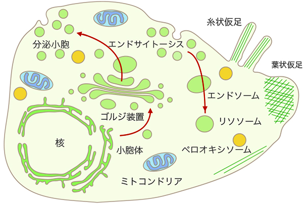 Fig. 1: Schematic diagram of the intracellular membrane system.
This system is connected by vesicular transport
Professor Satoh’s research group has paid particular attention to “membrane proteins” and has identified and analyzed factors involved in the “transport” process, the latter process of polarized transport, and is making progress in understanding where “sorting,” the former process, takes place.
The professor chose the Drosophila photoreceptor as the best model for this research. As only a small number of polarized transport researchers use this system, Professor Satoh independently developed most of the experimental systems.
“Since advanced molecular genetic tools are available in Drosophila, the combination of these genetic tools with sophisticated tissue analysis using an electron microscope or confocal laser microscope is the style of my research. To understand the mechanism of sorting processes, we need a system that can observe the sorting process with high temporal and spatial resolution. Therefore, we are currently trying to build such a system.”
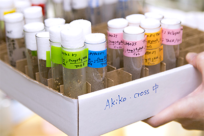
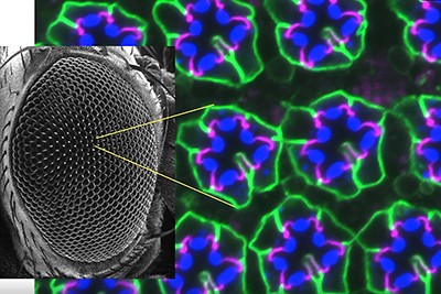 Fig. 2: Photoreceptors in Drosophila
Professor Satoh discovered a close relationship
between the Golgi apparatus and recycling endosomes.
This could be a breakthrough discovery
When asked what she finds interesting about her research, she replied as follows.
“I can find that there is a process that is quite different from what we have imagined. Through my research, I have come across various new phenomena that are not directly related to polarized transport. That is when my research gets the most interesting. Moreover, new directions of research arise from such unexpected discoveries. I can say that the best part of research is noticing and trying out new possibilities like that.”
The recently published research, “Discovery of a close relationship between two cell organelles,” was actually the result of such an “unexpected” discovery. There are three main points:
- Discovered that the Golgi apparatus, which is involved in the biosynthetic pathway(※1), and the recycling endosome (RE), which is involved in the recycling pathway(※2), repeatedly adhere and dissociate.
- Discovered that only limited types of biosynthesized membrane proteins are transported from the Golgi apparatus to the RE at the adhesion surface.
- Suggested that membrane protein sorting may take place on the adhesive surface of the Golgi apparatus and REs.
- ※1:The biosynthetic pathway is one of the vesicular transport pathways. It is a pathway in which membrane proteins and secretory proteins are synthesized in the endoplasmic reticulum, delivered to the Golgi apparatus, and then specifically transported from the Golgi apparatus to the plasma membrane and various organelles.
- ※2:The recycling pathway is one of the vesicular transport pathways. It is a pathway by which substances taken into a cell from the outside by endocytosis (eating and drinking) are transported back to the cell membrane.
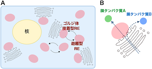 Fig. 3: Relationship between Golgi stacks (shown in gray) and REs (shown in pink)
A:A single cell contains many Golgi stacks and REs. There are two types of REs: REs that attach to the Golgi stacks and free REs, which repeatedly attach to and dissociate from the Golgi stacks. The REs also repeatedly attach to and dissociate from each other.
B:Only a portion of the membrane proteins transported from the endoplasmic reticulum to the Golgi stacks are transported to the plasma membrane via REs that attach to the Golgi stacks. Here membrane protein A, shown in green, is transported to the plasma membrane via the RE, whereas membrane protein B, shown in blue, is transported without going through the RE.
“In simple terms, we found that recycling endosomes and the Golgi apparatus performed polarized transport of membrane proteins by repeatedly attaching and dissociating.”
In addition, she made one major suggestion in this paper.
“The trans-Golgi network (TGN) at the end of the Golgi is thought to be part of the Golgi, but we think that the TGN is actually a RE (recycling endosome). It was common knowledge that TGN originally remained attached while RE was not. So, the two are called TGN when they are attached and RE when they are separated. Yet, we have found a possibility that the two may be identical organelles.”
When this research progresses, it may be a major discovery that will overturn previous concepts.
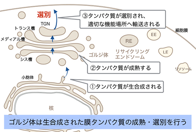 Fig. 4-1: Role of the Golgi apparatus and RE, part 1
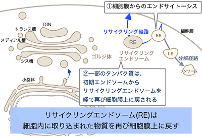 Fig. 4-2: Role of the Golgi apparatus and RE, part 2
Constantly engage in challenging research.
A goal is to present something new and conceptual through my research
Professor Satoh is enthusiastic about “continuing to conduct challenging research.”
“I am always willing to take on new challenges in my research, such as creating new systems and adopting new ways of thinking. My challenges will naturally involve my students. I would also like to create an environment where the students can enjoy their research while making mistakes.”
Recently, she has been conducting experiments using cultured human cells to test the generality of the unexpected organelle properties discovered in Drosophila. She seems satisfied with the research environment at Hiroshima University, saying, “There is a good access to confocal laser (scanning) microscope called FV3000, and I have the opportunity to use my own FV1000 and a super-resolution, high-speed microscope at RIKEN in Tokyo.”
Her main goal is to continue her challenging research and “present new concepts and ideas about the nature of organelles and their universality across species.”
 Opened on April 26, 2022

